Abstract
Hyalocytes in the pecten oculi and ciliary body of adult chickens and their response to Escherichia coli were investigated by transmission and scanning electron microscopy and the inflammatory response following the intravitreous injection of colloidal carbon examined by microscopy. In normal chickens, the hyalocytes were mainly found on the pleats of the pecten oculi and on the ciliary body. There were no hyalocytes on the retina. There is thus a close relationship between the vasculature in the tissues surrounding the vitreous chamber and the distribution of hyalocytes. The hyalocytes, which were predominantly spindle shaped or oval in contour, displayed a ruffled surface with occasional blebs, filopodia and lamellipodia. Flattened hyalocytes with relatively few and short pseudopodia were frequently observed, especially on the ciliary body. Hyalocytes responded quickly to E. coli bacteria which they phagocytosed. The response to colloidal carbon in the vitreous chamber had 3 distinct changes. In the 1st (2 d after carbon injection), the hyalocytes, the resident macrophages, actively ingested the carbon particles without significant leucocyte recruitment. In the 2nd stage (at 7-14 d), a large number of macrophages infiltrated the ciliary body and emigrated into the vitreous chamber. In the 3rd stage (at 30 d), the infiltration by macrophages into the ciliary body was complete. The carbon-laden macrophages disappeared from the vitreous body but accumulated on the pecten oculi and retina. They were exclusively drained through the scleral venous sinus in the iridocorneal angle.
Full text
PDF
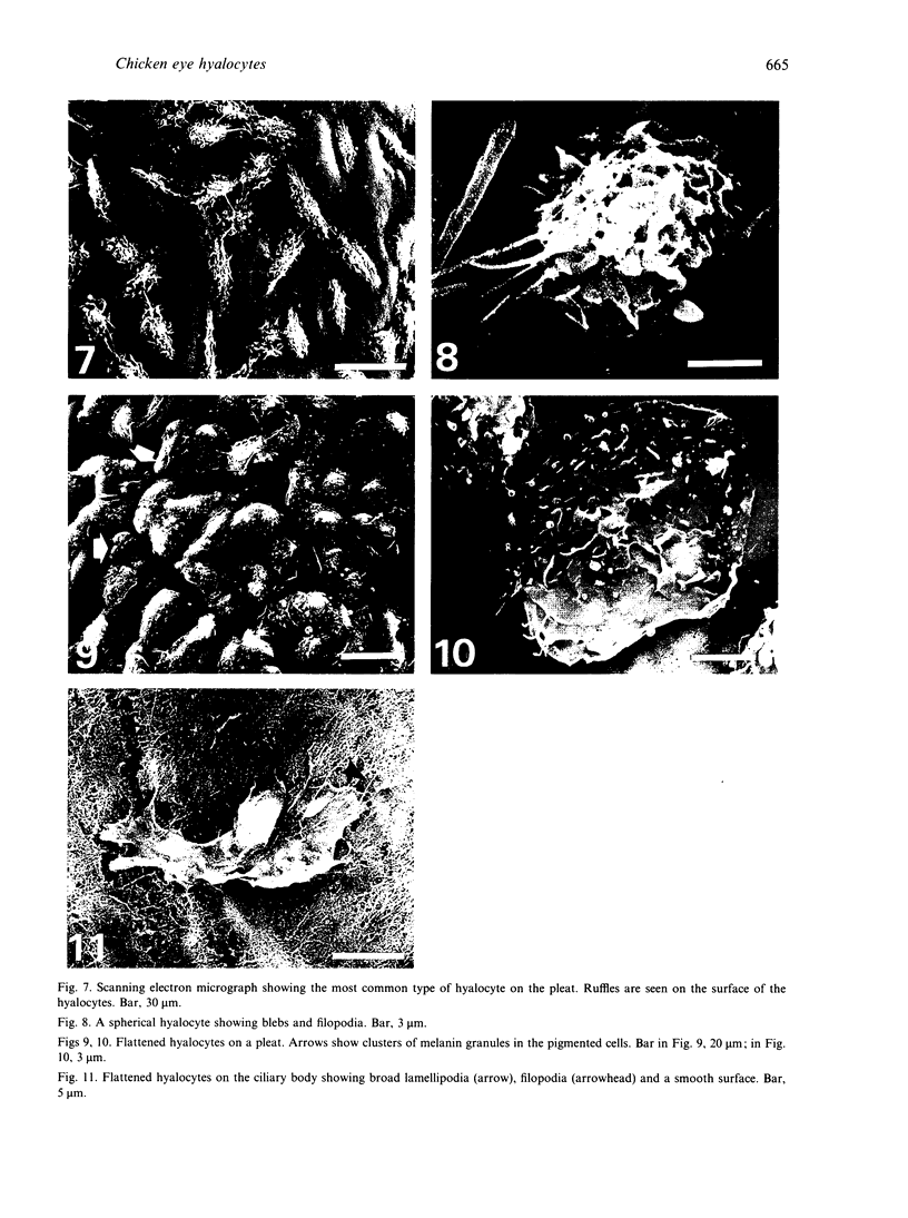
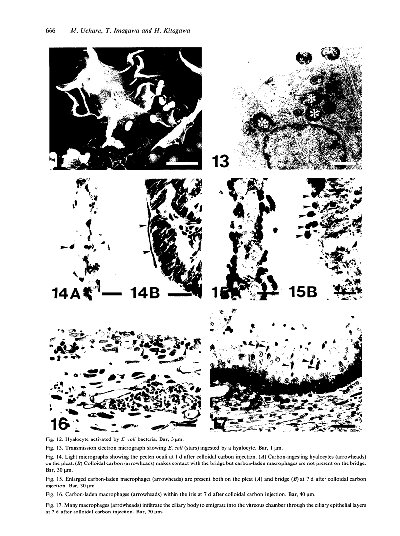
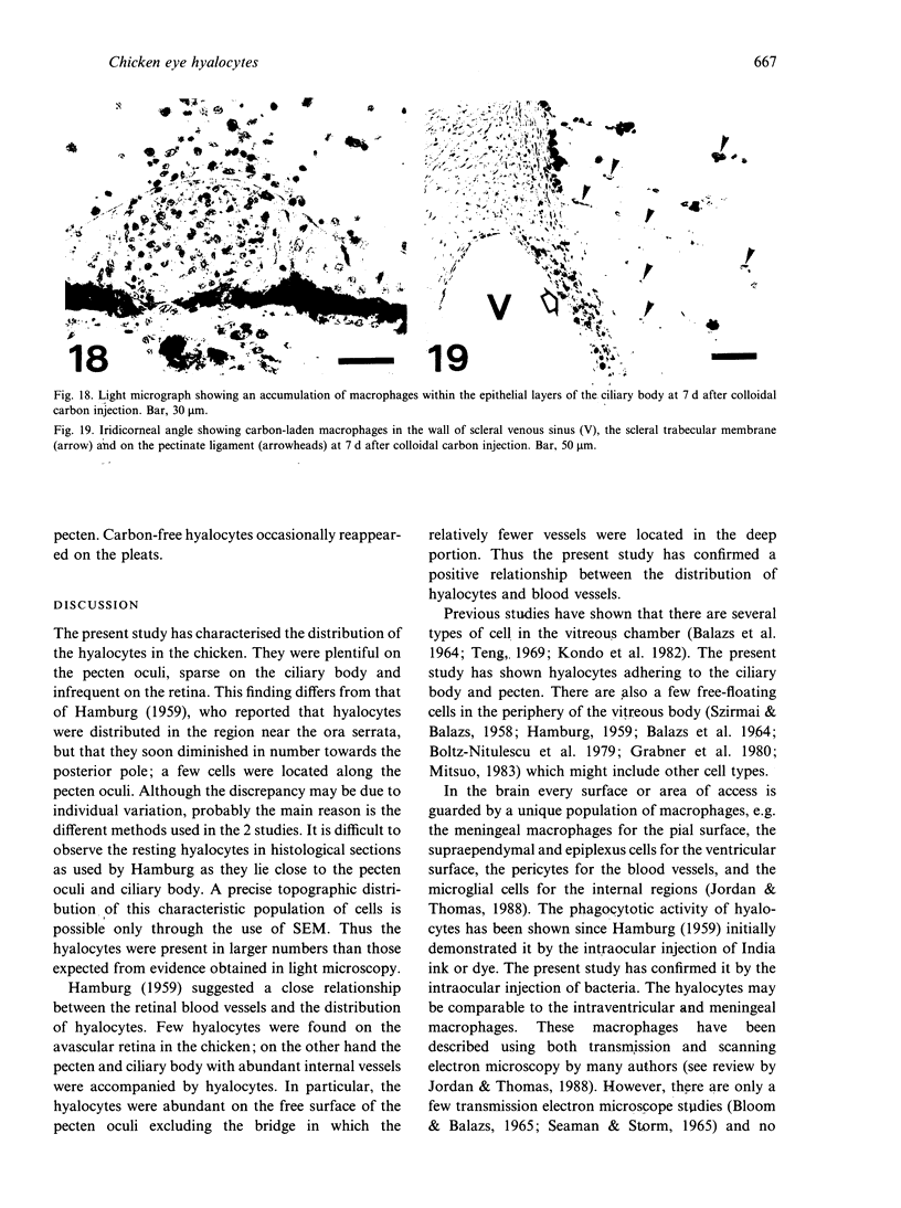
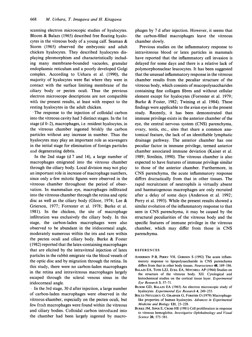
Images in this article
Selected References
These references are in PubMed. This may not be the complete list of references from this article.
- Andersson P. B., Perry V. H., Gordon S. The acute inflammatory response to lipopolysaccharide in CNS parenchyma differs from that in other body tissues. Neuroscience. 1992;48(1):169–186. doi: 10.1016/0306-4522(92)90347-5. [DOI] [PubMed] [Google Scholar]
- BALAZS E. A., TOTH L. Z., ECKL E. A., MITCHELL A. P. STUDIES ON THE STRUCTURE OF THE VITREOUS BODY. XII. CYTOLOGICAL AND HISTOCHEMICAL STUDIES ON THE CORTICAL TISSUE LAYER. Exp Eye Res. 1964 Mar;3:57–71. doi: 10.1016/s0014-4835(64)80008-7. [DOI] [PubMed] [Google Scholar]
- Bloom G. D., Balazs E. A. An electron microscopic study of hyalocytes. Exp Eye Res. 1965 Sep;4(3):249–255. doi: 10.1016/s0014-4835(65)80038-0. [DOI] [PubMed] [Google Scholar]
- Boltz-Nitulescu G., Grabner G., Förster O. Macrophage-like properties of human hyalocytes. Adv Exp Med Biol. 1979;121B:223–228. doi: 10.1007/978-1-4684-8914-9_20. [DOI] [PubMed] [Google Scholar]
- Burke J. M., Foster S. J. Ultrastructural demonstration of transretinal migration by vitreal macrophages marked with latex particles. Ophthalmic Res. 1982;14(5):317–325. doi: 10.1159/000265209. [DOI] [PubMed] [Google Scholar]
- Burke J. M., Sipos E., Cross H. E. Cell proliferation in response to vitreous hemoglobin. Invest Ophthalmol Vis Sci. 1981 May;20(5):575–581. [PubMed] [Google Scholar]
- Forrester J. V., Grierson I., Lee W. R. The pathology of vitreous hemorrhage. II. Ultrastructure. Arch Ophthalmol. 1979 Dec;97(12):2368–2374. doi: 10.1001/archopht.1979.01020020584018. [DOI] [PubMed] [Google Scholar]
- Forrester J. V., Lee W. R., Williamson J. The pathology of vitreous hemorrhage. I. Gross and histological appearances. Arch Ophthalmol. 1978 Apr;96(4):703–710. doi: 10.1001/archopht.1978.03910050393019. [DOI] [PubMed] [Google Scholar]
- Gloor B. P. On the question of the origin of macrophages in the retina and the vitreous following photocoagulation (autoradiographic investigations by means of 3H-thymidine). Albrecht Von Graefes Arch Klin Exp Ophthalmol. 1974 May 17;190(3):183–194. doi: 10.1007/BF00407092. [DOI] [PubMed] [Google Scholar]
- Grabner G., Boltz G., Förster O. Macrophage-like properaties of human hyalocytes. Invest Ophthalmol Vis Sci. 1980 Apr;19(4):333–340. [PubMed] [Google Scholar]
- HAMBURG A. Some investigations on the cells of the vitreous body. Ophthalmologica. 1959 Aug;138:81–107. doi: 10.1159/000303618. [DOI] [PubMed] [Google Scholar]
- Jordan F. L., Thomas W. E. Brain macrophages: questions of origin and interrelationship. Brain Res. 1988 Apr-Jun;472(2):165–178. doi: 10.1016/0165-0173(88)90019-7. [DOI] [PubMed] [Google Scholar]
- Kaiser C. J., Ksander B. R., Streilein J. W. Inhibition of lymphocyte proliferation by aqueous humor. Reg Immunol. 1989 Jan-Feb;2(1):42–49. [PubMed] [Google Scholar]
- Kaur C., Ling E. A., Gopalakrishnakone P., Wong W. C. Response of intraventricular macrophages to crotoxin-coated microcarrier beads injected into the lateral ventricle of postnatal rats. J Anat. 1990 Feb;168:63–72. [PMC free article] [PubMed] [Google Scholar]
- Lee W. R., Grierson I. Macrophage infiltration in the human retina. Albrecht Von Graefes Arch Klin Exp Ophthalmol. 1977 Sep 28;203(3-4):293–309. doi: 10.1007/BF00409835. [DOI] [PubMed] [Google Scholar]
- Ling E. A., Gopalakrishnakone P., Tan C. K. Electron-microscopical study of the choroid plexus and epiplexus cells in cats following a cisternal injection of crotoxin complex. Acta Anat (Basel) 1988;131(3):241–248. doi: 10.1159/000146523. [DOI] [PubMed] [Google Scholar]
- Lu J., Kaur C., Ling E. A. Immunophenotypic features of epiplexus cells and their response to interferon gamma injected intraperitoneally in postnatal rats. J Anat. 1994 Aug;185(Pt 1):75–84. [PMC free article] [PubMed] [Google Scholar]
- Lu J., Kaur C., Ling E. A. Intraventricular macrophages in the lateral ventricles with special reference to epiplexus cells: a quantitative analysis and their uptake of fluorescent tracer injected intraperitoneally in rats of different ages. J Anat. 1993 Oct;183(Pt 2):405–414. [PMC free article] [PubMed] [Google Scholar]
- Lu J., Kaur C., Ling E. A. Uptake of tracer by the epiplexus cells via the choroid plexus epithelium following an intravenous or intraperitoneal injection of horseradish peroxidase in rats. J Anat. 1993 Dec;183(Pt 3):609–617. [PMC free article] [PubMed] [Google Scholar]
- Mitsuo K. [Rabbit vitreous cells in tissue culture]. Nippon Ganka Gakkai Zasshi. 1983;87(12):1327–1336. [PubMed] [Google Scholar]
- Perry V. H., Andersson P. B., Gordon S. Macrophages and inflammation in the central nervous system. Trends Neurosci. 1993 Jul;16(7):268–273. doi: 10.1016/0166-2236(93)90180-t. [DOI] [PubMed] [Google Scholar]
- SEAMAN A. R., STORM H. K. ELECTRON MICROSCOPE OBSERVATIONS ON THE HYALOCYTES OF THE VITREOUS BODY IN THE DOMESTIC FOWL (GALLUS DOMESTICUS). Exp Eye Res. 1965 Mar;4:13–17. doi: 10.1016/s0014-4835(65)80003-3. [DOI] [PubMed] [Google Scholar]
- SZIRMAI J. A., BALAZS E. A. Studies on the structure of the vitreous body. III. Cells in the cortical layer. AMA Arch Ophthalmol. 1958 Jan;59(1):34–48. doi: 10.1001/archopht.1958.00940020058006. [DOI] [PubMed] [Google Scholar]
- Streilein J. W. Anterior chamber associated immune deviation: the privilege of immunity in the eye. Surv Ophthalmol. 1990 Jul-Aug;35(1):67–73. doi: 10.1016/0039-6257(90)90048-z. [DOI] [PubMed] [Google Scholar]
- Tagawa Y., Saga T., Matsuda H. [Morphological and histochemical studies on guinea pig hyalocytes]. Nippon Ganka Gakkai Zasshi. 1986 Mar;90(3):522–526. [PubMed] [Google Scholar]
- Teng C. C. An electron microscopic study of cells in the vitreous of the rabbit eye. II. Fibrocytes, plasma cells and an unidentified cell. Eye Ear Nose Throat Mon. 1969 Sep;48(9):532–539. [PubMed] [Google Scholar]
- Twining S., Potts D., Burke J. Acid proteases of vitreal macrophages. Curr Eye Res. 1984 Aug;3(8):1055–1062. doi: 10.3109/02713688409011752. [DOI] [PubMed] [Google Scholar]
- Uehara M., Oomori S., Kitagawa H., Ueshima T. The development of the pecten oculi in the chick. Nihon Juigaku Zasshi. 1990 Jun;52(3):503–512. doi: 10.1292/jvms1939.52.503. [DOI] [PubMed] [Google Scholar]


