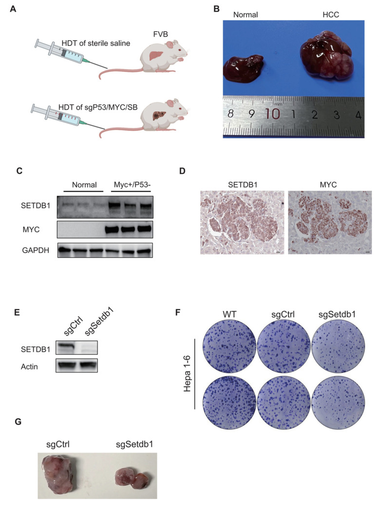
Figure 3.
In vivo and ex vivo validation of the SETDB1 function in HCC. (A) HCC model design. FVB mice were injected with normal saline or sgP53/c-Myc/SB plasmids, respectively. (B) Representative liver tissues of normal (left) and HCC mice (right). (C) The protein expression of SETDB1 and MYC in the normal and HCC tissues. (D) The IHC staining of SETDB1 and MYC in HCC liver tissues. (E) The SETDB1 was successfully knocked out in Hepa1-6 cells. (F) The SETDB1 knockout inhibited colony formation in Hepa1-6 cells. (G) The SETDB1 knockout suppressed tumor formation in the subcutaneous xenograft tumor model.

