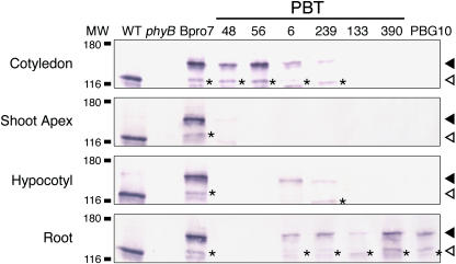
Figure 5.
Immunoblot Detection of phyB-GFP and Endogenous phyB in Cotyledons, the Shoot Apex, the Hypocotyls, and the Roots.
Proteins were extracted from 5-d-old seedlings grown under continuous white light (50 μmol m−2 s−1) and subjected to immunoblotting analysis with anti-Arabidopsis phyB antibody, mBA2. Closed and open arrowheads indicate positions of phyB-GFP and endogenous phyB, respectively. Asterisks indicate bands that are presumed to be a degradation product of phyB-GFP. MW, molecular weight (k). Each lane contained 20 μg of total proteins.

