Figure 2.
secGFP Labels ER and Post-ER Punctate Structures in the Presence of RAB-E1d[NI].
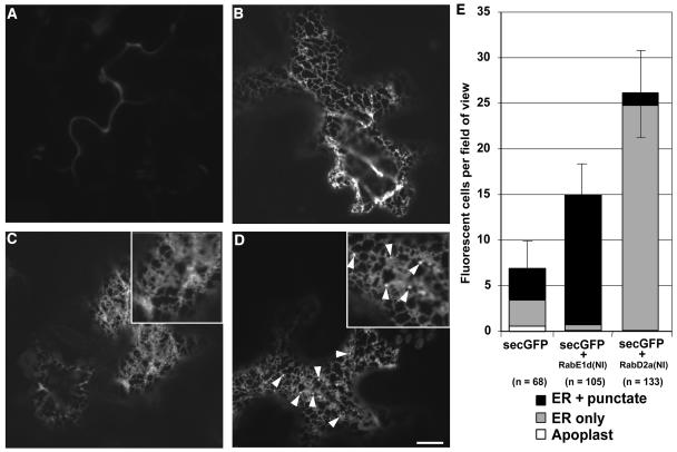
(A) to (D) Confocal images at high magnification of tobacco leaf epidermal cells expressing secGFP (A), GFP-HDEL (B), secGFP + RAB-D2a[NI] (C), and secGFP + RAB-E1d[NI] (D). Note the punctate structures indicated by arrowheads in the image (D) and the inset. Bar in (D) = 10 μm for (A) to (D).
(E) Average number of fluorescent cells and frequency of particular intracellular fluorescence patterns per field of view in cells expressing secGFP, secGFP + RAB-E1d[NI], and secGFP + RAB-D2a[NI]. n is the number of cells scored.

