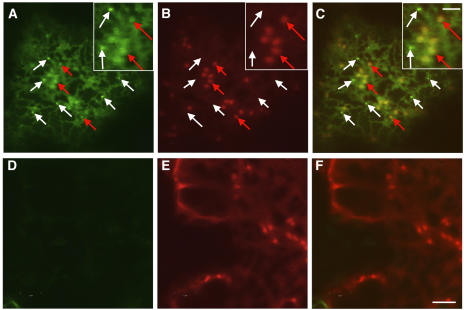
Figure 8.
YFP-Tagged RAB-E1d Does Not Label the PVC That Accumulates secGFP.
(A) to (C) Confocal images of a tobacco epidermal cell coexpressing secGFP and YFP-RAB-E1d at ∼50 h after infiltration, when secGFP accumulation is highest (Figure 1A). (C) is the merged image of secGFP fluorescence (A) and YFP-RAB-E1d fluorescence (B). White arrows in (A) to (C) and insets indicate examples of small, bright punctate structures that are labeled by secGFP but not by YFP-RAB-E1d. Red arrows indicate examples of large, faint Golgi punctate structures that are labeled by YFP-RAB-E1d fluorescence.
(D) to (F) Confocal images of a control tobacco epidermal cell expressing YFP-RAB-E1d without secGFP. The images were captured with identical image settings used for (A) to (C). (F) is merged from (D) and the YFP-RAB-E1d image (E). The absence of signal in the GFP image (D) excludes the possibility that the Golgi-sized faint punctate structures represent bleed-through of YFP fluorescence into the GFP detection channel. Bar in (F) = 5 μm for (A) to (F); bar in inset (C) = 2 μm for all insets.

