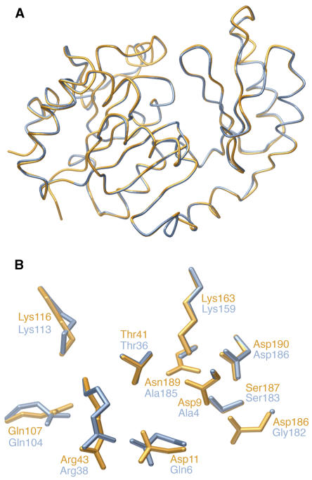
Figure 6.
Modeling of the SPP-Like Domain of SPS from Synechocystis sp PCC 6803.
(A) Superimposed view of the Synechocystis SPP in the closed conformation (orange) and the predicted structure of the SPP-like domain of the Synechocystis SPS (blue).
(B) Detailed view of the SPP active site in comparison with the homologous residues in the SPP-like domain of SPS.

