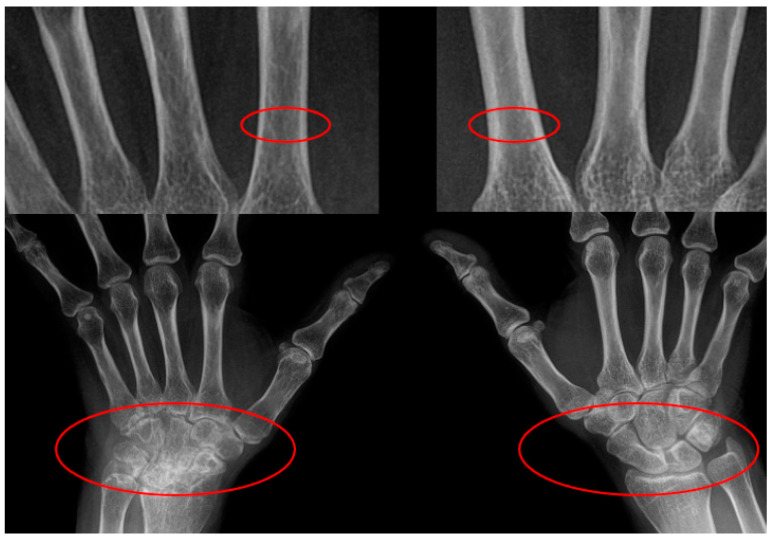
Figure 1.
Rheumatoid arthritis patients typically exhibit symmetrical arthritis, though some are known to show asymmetric arthritis. We encountered a patient with asymmetrical osteoporosis of the metacarpal bones and asymmetrical carpal lesions, which inspired this study. Upper Red Circled Regions: These focus on the cortical bone of the metacarpal diaphysis, showing differences in cortical thickness. The thinning of the cortical bone in these areas is more pronounced on one side, suggesting localized bone loss, often associated with periarticular osteoporosis in rheumatoid arthritis. Lower Red Circled Regions: These highlight the wrist joint areas, illustrating bone erosion, joint space narrowing, or other joint damage. A clear asymmetry between the left and right wrist joints is observed, indicating more severe joint damage on one side, likely due to localized inflammation and progressive joint damage.

