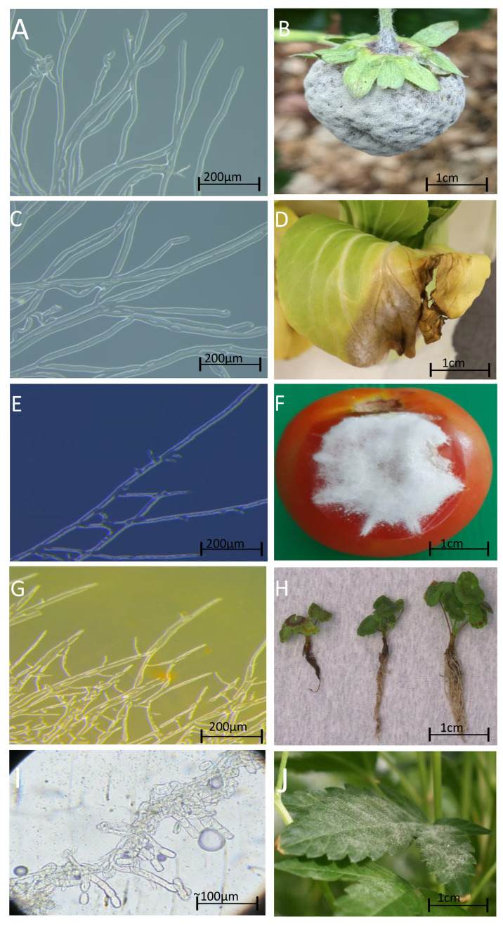
Figure 1.
Microscopic and macroscopic images of fungal pathogens detected in protected cropping environments. (A) Laboratory-grown Botrytis cinerea on a potato dextrose agar plate and viewed under a Leica M205 FA Stereomicroscope. (B) Strawberry fruit infected with Botrytis cinerea in field (photo taken by Marlo Molinaro La Trobe University). (C) Sclerotinia sclerotiorum grown in the laboratory on a potato dextrose agar plate and viewed under a Leica M205 FA Stereomicroscope. (D) Bok choy leaf infected with Sclerotinia sclerotiorum, 8 days after inoculation. (E) Laboratory grown putative Fusarium oxysporum collected from CEA, grown on a potato dextrose agar plate and viewed under a Leica M205 FA Stereomicroscope. (F) Fusarium growing on a tomato, reprinted with permission from [30]. Copyright 2021 Zahir Shah Safari from Leibniz Universität Hannover. (G) Laboratory grown Pythium irregulare now Globisporangium irregulare on a potato dextrose agar plate viewed under a Leica M205 FA Stereomicroscope, obtained from Dr. Niloofar Vaghefi from University of Melbourne. (H) Pythium irregularre now known as Globisporangium irregulare growing on geraniums taken and unchanged from CABI Plantwise Plus website (https://plantwiseplusknowledgebank.org/doi/10.1079/PWKB.Species.46152, accessed on 16 October 2024) under a Attribution-NonCommercial-NoDerivatives 4.0 International (CC BY-NC-ND 4.0) license taken by Michael Evans, University of Arkansas. (I) Suspected Golovinomyces cichoracearum grown on onion viewed under a microscope, taken from the Global Biodiversity Information Facility website (https://www.gbif.org/occurrence/4891810393, accessed on 16 October 2024) by Schmidt Dávid (licensed under http://creativecommons.org/licenses/by-nc/4.0/, accessed on 17 October 2024). Golovinomyces is an obligate fungus and therefore unable to be cultured in the laboratory on a growing media. (J) Suspected Golovinomyces cichoracearum grown on Cannabis sativa L. courtesy of industry partner. Scale = 200 µm on (A,C,E,G), 1 cm on (B,D,F,H,J) and ~100 µm for (I).

