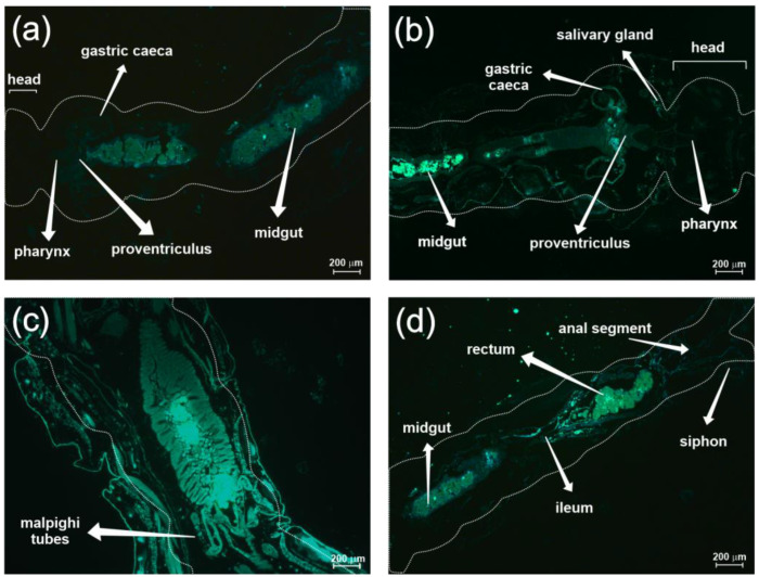
Figure 4.
Fluorescence tracing of chitosan nanoparticles in the digestive system of mosquito larvae. (a) Head, gastric caecum, midgut, proventriculus, and pharynx of larva treated with chitosan nanoparticle without β-myrcene (CNPP), at 5× magnification. (b) Head, salivary gland, caecum, midgut, proventriculus, and pharynx of larva treated with chitosan nanoparticle with β-myrcene (CNPM), at 5× magnification. (c) Midgut, rectum, midgut, ileum, anal segment, and siphon, of larva treated with CNPP, at 20× magnification. (d) Midgut of larva treated with CNPM, at 5× magnification. The dashed lines represent the exoskeleton of the larvae.

