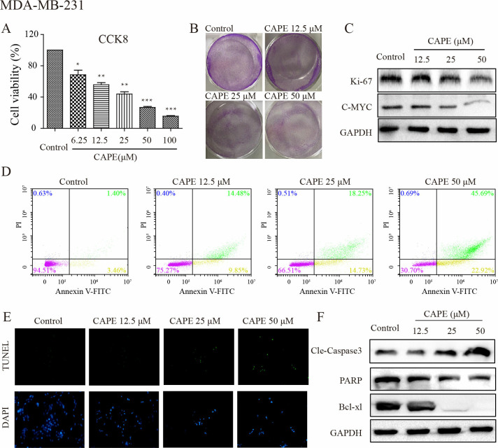
Fig 1. CAPE inhibited cell proliferation and induced cell apoptosis of MDA-MB-231 cells.
(A) Cells were treated with different concentrations of CAPE (6.25–100 μM) for 72 h, and CCK8 was used for cell viability detection. (B) Cell were treated with CAPE (12.5, 25, 50 μM) for 5 days to investigate cell colony formation. (C) The inhibition of cell proliferation was measured by western blot for the level of Ki-67 and C-MYC using CAPE (12.5, 25, 50 μM) treatment for 24 h. (D) Cells were treated with CAPE (12.5, 25, 50 μM) for 48 h, and the cell apoptosis was detected by TUNEL/DAPI dual staining. (E) Cells were treated with CAPE (12.5, 25, 50 μM) for 24 h, and western blot was used to detect the expression of Cle-Caspase3, PARP, and Bcl-xl. (F) Cells were treated with CAPE (12.5, 25, 50 μM) for 48 h, and apoptosis was determined by Annexin V-FITC/PI dual staining. Values represent the mean ± SD from three independent experiments; *p <0.05, **p<0.01, ***p<0.001: CAPE groups compared with the control group.

