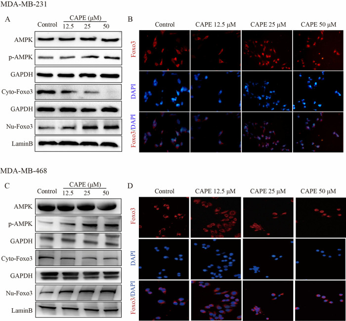
Fig 4. CAPE activated the AMPK and Foxo3 signals.
(A, C) MDA-MB-231 and MDA-MB-468 cells were treated with CAPE (12.5, 25, 50 μM) for 12 h, and the protein levels of p-AMPK, GAPDH, Foxo3 (respectively in the cytoplasm and nucleus), and LaminB were analyzed by western blot. (B, D) MDA-MB-231 and MDA-MB-468 cells were treated with CAPE (12.5, 25, 50 μM) for 12 h, and the nuclear translocation of Foxo3 was determined by immunofluorescence staining. Values represent the mean ± SD from three independent experiments.

