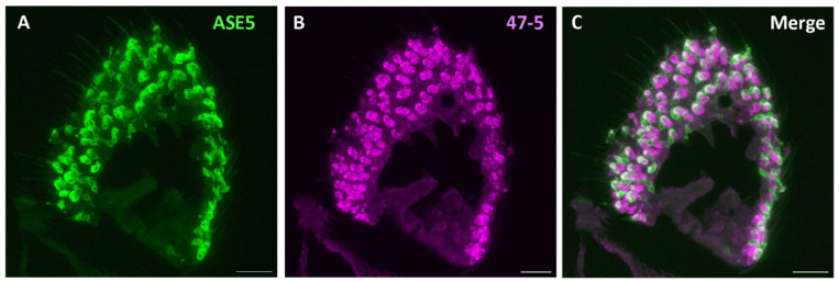
Figure 7.
Expression pattern of V-ATPase in the tormogen cells of D. melanogaster antennal cross-sections. (A) Immunofluorescence image of mCD8::GFP (anti-GFP, green) and V-ATPase (47-5, magenta). (B) From the antennal cross sections of ASE5-GAL4 fly line labeling tormogen cells. (C) Observation of major colocalization of mCD8::GFP and V-ATPase in tormogen cells. Scale bar: 17 µm.

