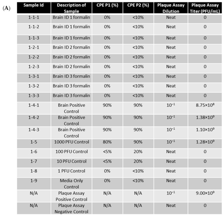
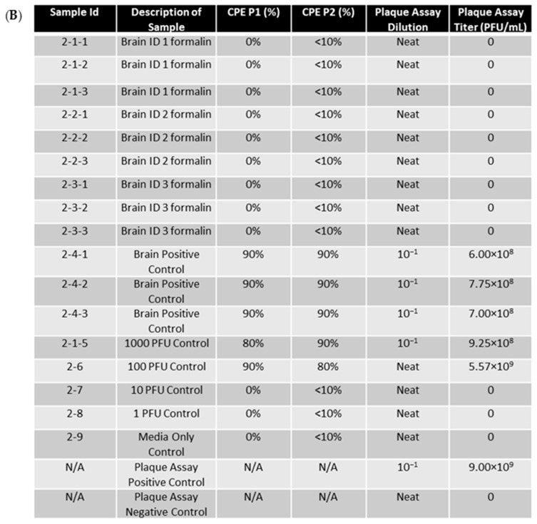
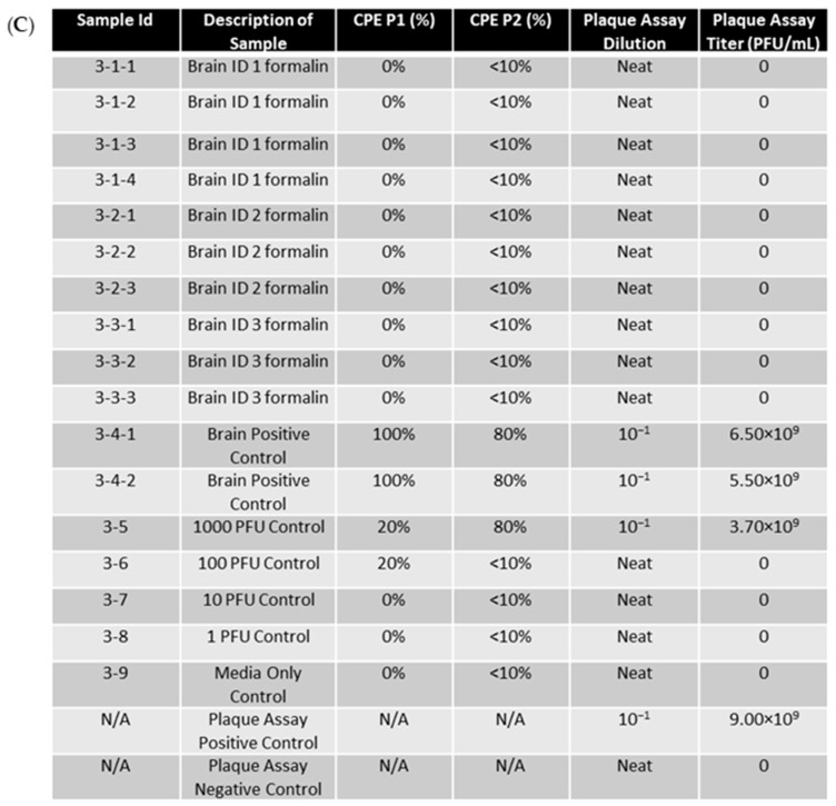
Figure 5.
EEEV-infected NHP brains were fixed in 10% formalin and homogenized alongside positive control tissue samples. RNA extraction was then performed on samples. RNA was then electroporated into BHK-21 cells. RNA samples that were more than 0.5 (electroporation volume) were split into separate samples (ex: 3-1 into 3-1-1, 3-1-2, and 3-1-3) for electroporation into cells. Cells were then placed in flasks for 72 h as a first passage. A second passage was also performed for 72 h. CPE was observed and recorded daily. PFU controls were created by adding diluted stock virus at the designated PFU directly to cell culture. Plaque assay was performed on RNA extracted from formalin-fixed samples and control samples to determine if genomic replication and infectious virus production were occurring. Stock virus was diluted and used as a plaque assay positive control. The limit of detection for the assay was 1 PFU/mL. (A) CPE and plaque assay data for replicate experiment 1. (B) CPE and plaque assay data for replicate experiment 2. (C) CPE and plaque assay data for replicate experiment 3.


