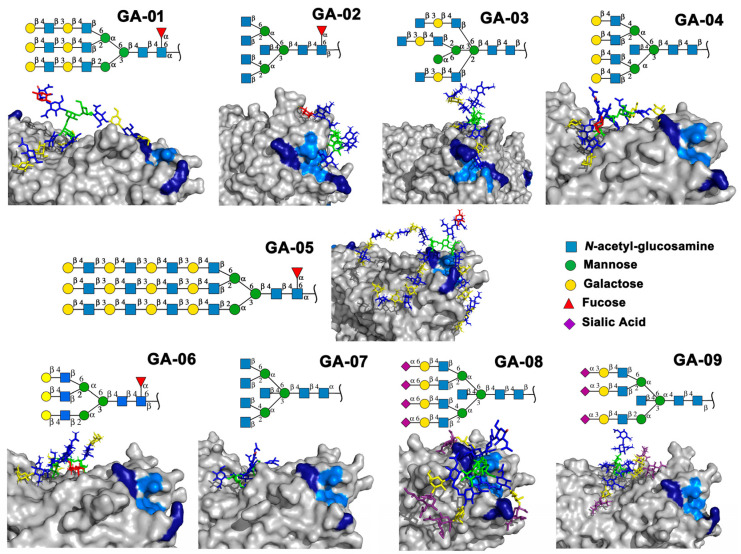
Figure 4.
Molecular docking of the experimental N-glycans from the Mammalian Glycan Array 5.4 with rTBL-1 structures. The graphical 2D representation of N-glycans is shown according to the Symbol Nomenclature for Glycans (SNFG) [28]. The N-glycans from the mammalian glycan array “Version 5.4” are labeled as GA-##, where ## is the number described by Martínez-Alarcón et al. (2020) [13] and is related to the signal’s relative intensity of lectin recognition. Docking results are shown as topological representations of interactions at the same surface location between the N-glycans with the recombinant lectins: rTBL-1, R104Q, and with the R131Q lectin. The N-glycan in topological representations is presented as sticks colored according to the SNFG (as indicated by geometric codes), the protein surface in gray, highlighting the CBP in light blue and the arginine residues in dark blue. GA, glycan array.

