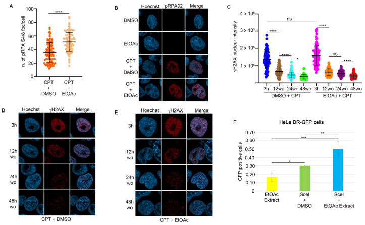
Figure 2.
EtOAc does not affect DNA damage repair ability induced by camptothecin. (A) RPA32 S4/8 foci intensity in HeLa cells treated for one hour with EtOAc at 0.63 μg/mL, followed by incubation with 1 μM CPT for an additional two hours. Foci number was measured with Fiji software. **** p < 0.0001. (B) Representative images of pRPA32 following incubation with the indicated reagents. All immunofluorescence images were acquired with the LSM900 using AiryScan2. Magnification 63×. (C) γH2AX intensity was measured in HeLa cells pre-treated with EtOAc extract or DMSO for one hour, followed by 1 μM CPT treatment. At the end of incubation time we performed a drugs washout, monitoring the DNA repair as a measure of γH2AX nuclear signal. Immunofluorescence images were analyzed with Fiji software. * p < 0.05 and **** p < 0.0001. (D) Representative images of H2AX S139 nuclear intensity in HeLa cells treated with 1 μM CPT and DMSO for three hours, followed by drug washout to monitor H2AX recovery. Magnification 63×. (E) Immunofluorescence analysis of HeLa cells treated for one hour with EtOAc at 0.63 μg/mL, followed by incubation with 1 μM CPT for an additional two hours. Magnification 63×. (F) HeLa cells stably transfected with the pDR-GFP vector were co-transfected with I-SceI endonuclease and incubated with 0.63 μg/mL of EtOAc extract for 24 h. HR activity was measured using FACS analysis, with GFP levels serving as an indicator of HR frequency. Data are presented as means ± standard deviation from three independent experiments. Statistically significant differences are indicated by * p < 0.05, ** p < 0.01, and *** p < 0.001.

