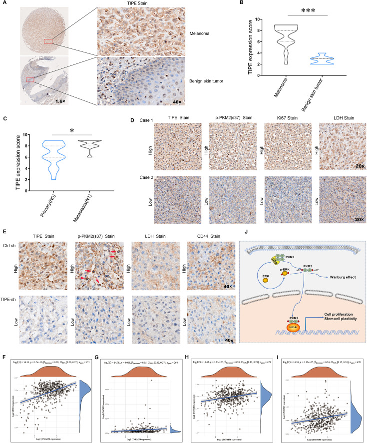
Figure 7. TIPE positively corelated with cancer stem cell (CSC) markers and the levels of p-PKM2(Ser37).
(A, B) Higher expression of TIPE was observed in melanoma tumor tissues than in the control, as evidenced by immunohistochemistry. (C, D) The expression of TIPE correlated well with p-PKM2(Ser37) in melanoma tumor tissues. (E) Similarly, a good correlation was observed between TIPE, p-PKM2(Ser37), LDH, and CD44 in mouse xenografts. (F–I) In addition, the expression of TIPE was positively correlated with CSCs markers, including BMI1, NANOG, NOTCH1, and POU5F1 in TCGA dataset. (J) A brief model depicting the functional impact of TIPE on metabolic reprogramming in melanoma. ***p < 0.001. The data represent the means ± SEM of three replicates "*p<0.05.


