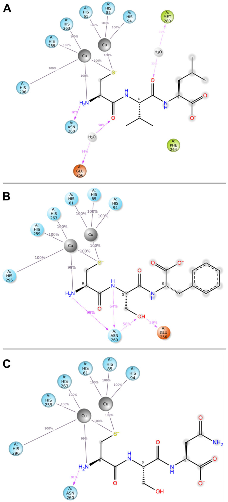
Figure 5.
Interactions between ligand atoms and tyrosinase residues for CVL (A), CSF (B), and CSN (C) tripeptides (schematic). The contacts shown occurred >30% of the simulation time. The colors of enzyme residues correspond to their chemical nature: polar (blue); negatively charged (orange); and hydrophobic (green). Copper atoms are represented as gray spheres. Percent values indicate the frequency of the interaction during analyses.

