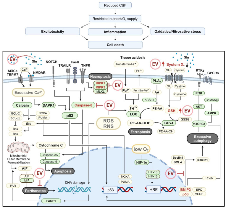
Figure 2.
Schematic overview of brain cell death in ischemic stroke. Reduced cerebral blood flow (CBF) leads to decreased nutrient and oxygen supply, triggering excitotoxicity, inflammation, and oxidative/nitrosative stress, which ultimately result in brain cell death. The molecular mechanisms underlying various forms of brain cell death in ischemic stroke are illustrated in the figure. Excitotoxicity-induced receptor activation and intracellular calcium overload contribute to autophagy-related cell death and apoptosis via DAPK1, CaMKKβ, and calpain activation, while also triggering ferroptosis through PLA2 activation, which provides arachidonic acid (AA). Oxidative/nitrosative stress induces mitochondrial outer membrane permeabilization, DNA damage, and lipid peroxidation, driving apoptosis, parthanatos, and ferroptosis. Furthermore, external signals, including proinflammatory cytokines, induce apoptosis and autophagy-related cell death through MAPK and NOTCH signaling, caspase-8 activation, and mTORC1 inhibition. Under the conditions of ATP depletion, rather than activating caspase-8, cell death signaling induced by TNF-α and FasL promotes necroptosis by forming a necrosome complex comprising RIPK1/3 and MLKL. Key molecules or pathways targeted by the extracellular vesicles (EV) are highlighted in red color. Green boxes indicate molecules with enzymatic functions. Schematic illustration was created with BioRender.com.

