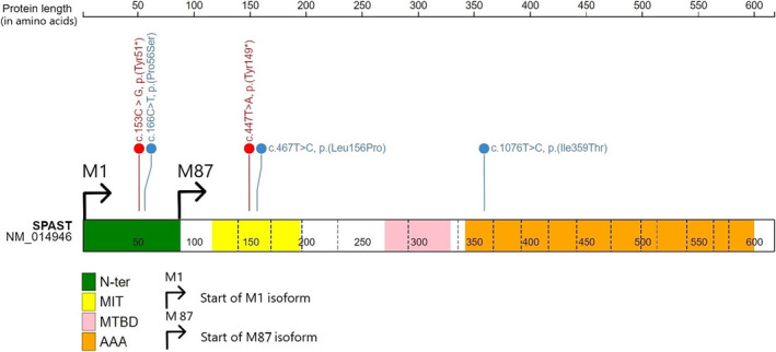
FIGURE 2.

Diagrammatic representation of the SPAST gene and the localization of variants relative to the protein's functional domains of interest. The different functional domains are represented with colours: In green the N‐ter domain (N‐ter), in yellow the Microtubule Interacting and Trafficking domain (MIT), in pink the Microtubules binding domain (MTBD) and orange the AAA domain (AAA). The five different SNVs are represented according to their position in SPAST, with missense variants in blue and nonsense variants in red. The N‐ter domain, in green, is only present in the M1‐isoform of the protein.
