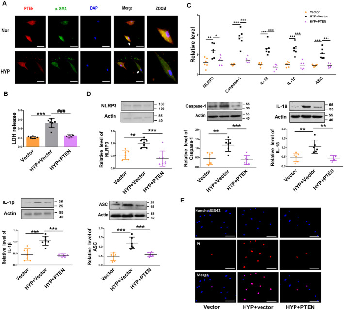
Figure 4. PTEN overexpression inhibits PASMCs pyroptosis.

A, Immunofluorescence assay of PTEN in hypoxic PASMCs. The cells were stained for PTEN (red) and α‐smooth muscle actin (green), and nuclei were stained with DAPI (blue). Scale bars=100 μm. B, PTEN overexpression reversed the increased LDH activity induced by the exposure of PASMCs to hypoxia for 48 h, as indicated by LDH analysis (Vector, n=6; HYP+Vector, n=6; HYP+PTEN, n=6). C, D, Overexpression of PTEN reversed the increased protein and mRNA levels of NLRP3, caspase‐1, interleukin‐1β, interleukin‐18, and ASC induced by the exposure of PASMCs to hypoxia for 48 h, as detected by qRT‐PCR and western blot, respectively (Vector, n=6–7; HYP+Vector, n=6–7; HYP+PTEN, n=6–7). E, PTEN overexpression reversed the increased positive PI staining induced by the exposure of PASMCs to hypoxia for 48 h. Images of fluorescence staining with PI (red) and Hoechst 33342 (blue). Scale bars=100 μm. Each data point in the figure represents a unique biological replicate. Statistical analysis was performed with one‐way ANOVA. The data are presented as the mean±SD. *P<0.05, **P<0.01, ***P<0.001. ASC indicates apoptosis‐associated speck‐like protein containing a caspase recruitment domain; HYP, hypoxia; LDH, lactate dehydrogenase; NLRP3, nucleotide‐binding oligomerization segment‐like receptor family 3; PASMC, pulmonary artery smooth muscle cell; PI, propidium iodide; and PTEN, phosphate and tension homology deleted on chromosome 10.
