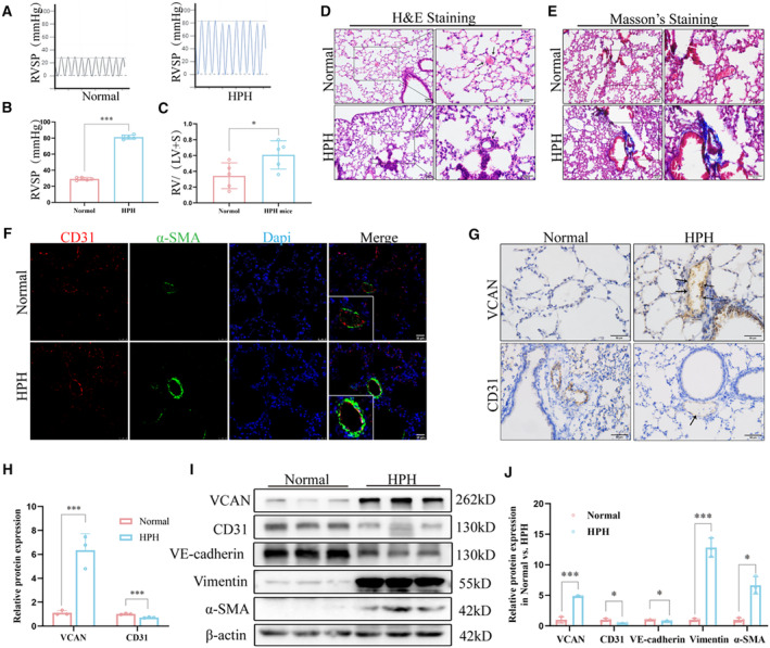
Figure 2. Vascular remodeling, EndMT, and increased VCAN in pulmonary vessels of successfully constructed HPH mouse models.

A and B, RVSP was measured using pressure sensors in the normoxia and hypoxia groups (n=5). ***P<0.001. (C) RV/(LV+S) of heart in mice of normoxia and hypoxia groups (n=5). *P<0.05. (D and E) H&E staining and Masson's trichrome staining of representative lung sections from normoxia and hypoxia groups. Arrows show normal or thickened blood vessels. Blue indicates collagen deposition (n=3). ×200, scale bar: 50 μm; ×400, Scale bar: 50 μm. (F) Co‐expression of CD31 (red) and α‐SMA(green) in the pulmonary arteries of HPH mice (n=3). ×400, scale bar: 25 μm. (G and H) Representative images of immunohistochemical staining showed that VCAN was upregulated in the pulmonary endothelium of HPH mice compared with the normal group. Positive staining was indicated by a brown color and pointed out with black arrow (n=3). ×400, scale bar: 50 μm. ***P<0.001. I and J, The protein levels and densitometric quantification of VCAN, CD31,VE‐cadherin, vimentin, and α‐SMA were determined by Western blot analysis after the hypoxia treatment (n=3). *P<0.05, ***P<0.001. An unpaired 2‐tailed Student t test was performed for comparisons between 2 groups. CD31 indicates platelet endothelial cell adhesion molecule‐1; EndMT, endothelial‐to‐mesenchymal transition; H&E, hematoxylin–eosin; HPH, hypoxia‐induced hypertension; α‐SMA, α‐smooth muscle Actin; RVSP, right ventricular systolic pressure; VCAN, versican; and VE‐cadherin, vascular endothelial cadherin.
