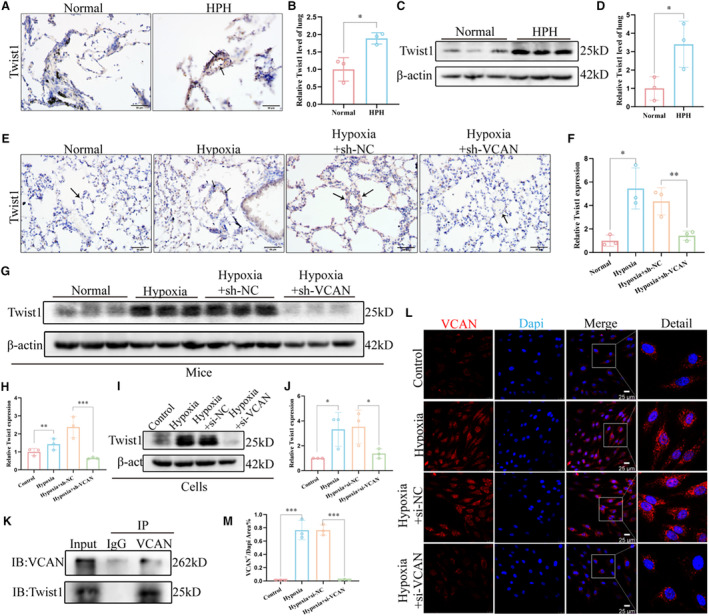
Figure 5. VCAN promoted EndMT via targeting transcription factor Twist1.

A and B, Representative images of immunohistochemical staining showed that Twist1 was upregulated in the pulmonary endothelium of patients with HPH compared with the normal group (n=3). Positive staining was indicated by a brown color and pointed out with black arrows. ×400, scale bar: 50 μm. *P<0.05. (C and D) The protein level and densitometric quantification of Twist1 in lung tissues of 2 groups were determined by Western blot analysis (n=3). *P<0.05. (E and F) Representative images of immunohistochemical staining showed that knocking down VCAN in vivo reduces upregulated Twist1 in the pulmonary vascular endothelium of HPH mice. Positive staining is indicated by a brown color and pointed out with black arrows (n=3). ×400, scale bar: 50 μm. *P<0.05, **P<0.01. (G–J) The protein levels and densitometric quantification of Twsit1 in vivo and in vitro knockdown VCAN assays were determined by Western blot analysis (n=3). *P<0.05, **P<0.01, ***P<0.001. K, The CO‐IP experiment showed that VCAN and twist1 can bind to each other in HPAECs. The target protein VCAN was immunoprecipitated with either anti‐Twist1 antibody or IgG. L, Enlarged images of the immunofluorescence staining of HPAECs nucleus. × 400, scale bar: 25 μm. An unpaired 2‐tailed Student t test was performed for comparisons between the normal group and patients with HPH, while ANOVA with the Tukey test was performed for comparison between 4 groups of in vivo and in vitro experiments. CO‐IP indicates Co‐Immunoprecipitation; EndMT, endothelial‐to‐mesenchymal transition; HPAECs, human pulmonary artery endothelial cells; HPH, hypoxia‐induced pulmonary hypertension; and VCAN, versican.
