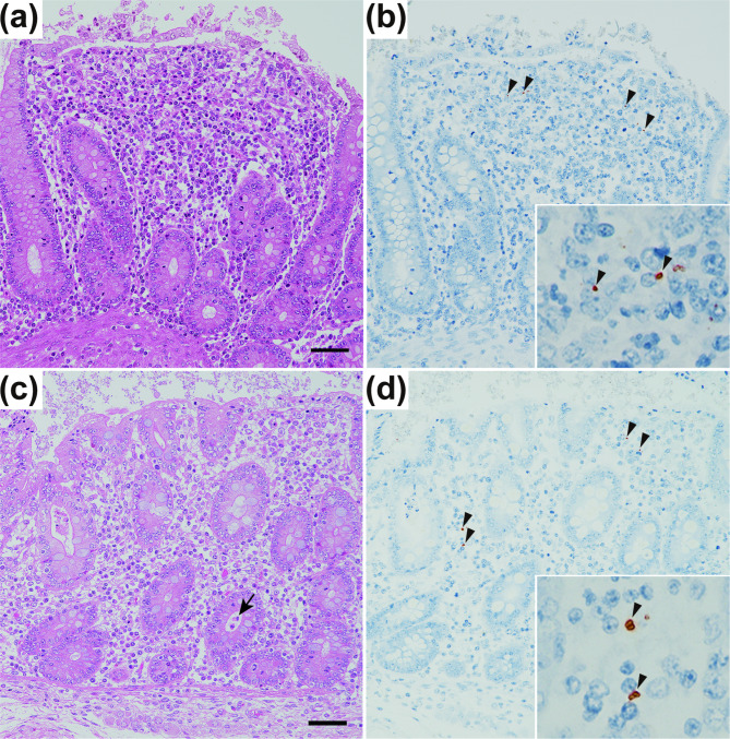
Fig. 4.
Histopathology of the cecum of pigs 7 days postadministration of the Salmonella Typhimurium L-3569 (a,b) and L-3569TC (c,d) strains. The lamina propria was expanded and contained many neutrophils, macrophages, lymphocytes, and plasma cells (a,c). Occasionally, small cryptic abscesses were observed (c; arrow). Hematoxylin and eosin stains; bar = 50 μm. Immunohistochemically, Salmonella O4 antigen-positive bacteria (b,d; arrowheads in the insert) were observed in the lamina propria by the antibody-labeled polymer method.

