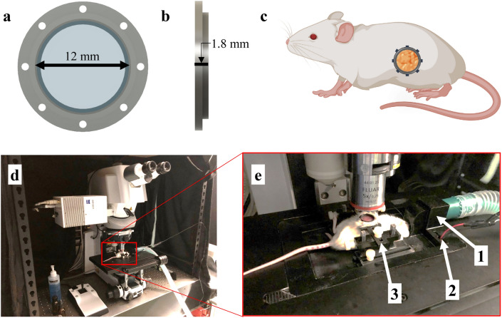
Fig. 1.
Surgically implanted abdominal imaging window allows longitudinal intravital imaging of pancreatic tumors in vivo. (a,b) Computer-aided design (CAD) of an abdominal pancreatic imaging window (PIW) frame holding a circular glass coverslip (12 mm diameter). (c) Diagram of a mouse (using BioRender.com) indicating the anatomical location of the surgically implanted PIW. The PIW was 3D-printed with biocompatible, acrylonitrile butadiene styrene (ABS) plastic and surgically implanted over the tumor site, providing longitudinal imaging access to the pancreas in vivo. (d) Photo of laser-scanning confocal/multiphoton microscope with (e) a custom, 3D-printed stage insert to stabilize the animal for anesthesia and imaging. The stage is equipped with (1) a gas anesthesia port, (2) an electrical heating element and (3) a holder to minimize motion artifacts during imaging. This figure was originally published by Samuel et al.39.

