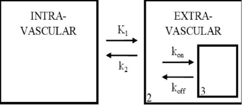
Figure 1.

Three-compartment model used in analysis of the brain tissue. Compartments 1, 2 and 3 represent the possible environments for the radioligand. Interactions between compartments are governed by variables for diffusion, and binding kinetics (kon, koff). This figure is a modified version of the one reported by Mintun et.al (1984) (1)
