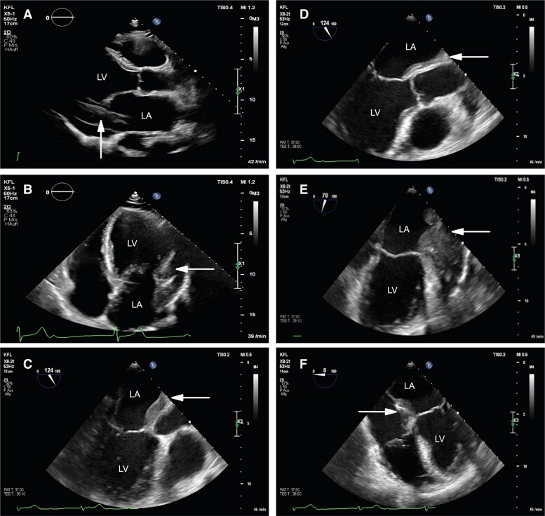
Figure 2.
Echocardiography. (A and B) Transthoracic echocardiography suspecting thickening of left heart and pericardial effusion in (A) parasternal long-axis view and in (B) apical four-chamber view (white arrow). (C–F) Transoesophageal echocardiography suggesting infiltrative character in (C) mid-oesophageal long-axis view, in (D) mid-oesophageal aortic valve long-axis view, in (E) two-chamber view and (F) four-chamber view (white arrow). LA, left atrium; LV, left ventricle.

