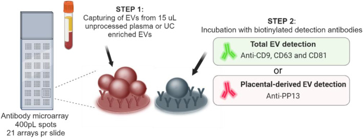
FIGURE 2.

Principle of analysis of the extracellular vesicle (EV) Array. A customized antibody microarray is generated onto glass slides. EVs from unprocessed plasma or UC enriched EVs are added and EVs are captured during incubation. Step 2 is either performed using a cocktail of anti‐CD9, CD63 and CD81 to detect the total amount of EVs or by using anti‐PP13 to detect only placenta‐derived EVs. After Step 2 fluorescent streptavidin is added prior to scanning.
