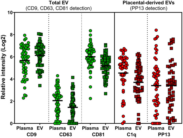
FIGURE 4.

Comparing results from raw plasma extracellular vesicles (EVs) vs ultracentrifuged pelleted enriched EVs. Green dots to the right 3 columns indicate detection of total EVs using antibodies to CD9, CD63 and CD81 as specific tetraspanins markers of EVs. Red dots at the right three columns marks the placental‐derived EVs detected by anti‐PP13 antibodies. The couple of samples plotted in each shows the signal obtained from the raw plasma preparation vs the pelleted EV sample indicating the reliability and accuracy of the direct plasma samples overlay on the surface of the glass slide of the EV Array.
