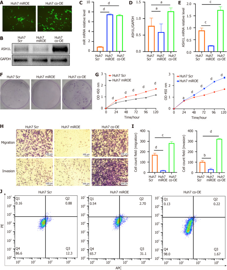
Figure 7.
The expression of ASH1L and proliferation assay, migration and invasion assay in Huh7 scrambled, microRNA overexpression and co-overexpression cell lines. A: Fluorescence imaging was conducted to visualize the transfection efficiency of lentivirus in Huh7 cells, allowing for the assessment of viral uptake and distribution within the cells; B: Western blot analysis was performed on Huh7 cells to evaluate protein expression levels among the group of scrambled (Scr), microRNA over-expression (miROE), and microRNA and ASH1L over-expression (co-OE) groups, providing insights into the molecular effects of these genetic modifications; C: Quantitative real-time polymerase chain reaction (RT-qPCR) analysis was carried out to quantify the expression of specific genes at the transcriptional level in Huh7 Scr, miROE, and co-OE groups, offering a detailed view of changes in gene expression post-transfection; D: The quantitative results of the western blot analysis provided numerical data on protein expression differences between the Huh7 Scr, miROE, and co-OE groups, allowing for statistical comparison of the effects of lentiviral transfection; E: RT-qPCR analysis was also conducted to compare the relative gene expression levels in Huh7 Scr, miROE, and co-OE groups, further validating the molecular changes induced by the transfections; F: A colony formation assay was used to assess the proliferative capacity of Huh7 Scr, miROE, and co-OE groups, indicating the clonogenic potential of the cells after genetic manipulation; G: Cell counting kit 8 assays were implemented to measure the cell proliferation rate in Huh7 Scr, miROE, and co-OE groups, providing a quantitative measure of cell growth and viability; H: Transwell chamber tests were conducted to determine the migratory and invasive capabilities of Huh7 Scr, miROE, and co-OE groups, revealing the functional consequences of altered gene expression on cell behavior; I: Quantitative analyses of the Transwell chamber test results provided statistical evaluation of the differences in cell migration and invasion among Huh7 Scr, miROE, and co-OE groups, quantifying the impact of the genetic interventions on these cellular processes; J: Cell apoptosis was detected by flow cytometry, and the results showed the influence of miR-142-3p and ASH1L overexpression on apoptosis. aP < 0.05. bP < 0.01. cP < 0.001. dP < 0.0001. GAPDH: Glyceraldehyde-3-phosphate dehydrogenase; Scr: Scrambled; miROE: MicroRNA over-expression; co-OE: MicroRNA and ASH1L over-expression; APC: Allophycocyanin conjugate; PE: Phycoerythrin.

