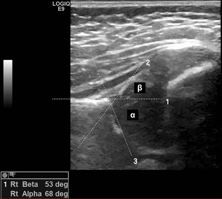
Figure 1. Hip ultrasound image.
The image shows the correct measurement of alpha and beta angles.
Ensuring the accuracy of an image involves two crucial steps. The first step is identifying eight anatomical landmarks on the image: the chondro-osseous border, femoral head, synovial fold, joint capsule, labrum, the cartilage portion of the roof, the bony portion of the roof, the bony rim (also known as the turning point between the bony roof's concavity and convexity), and the ilium.
The second step is completing the usability checklist, which ensures the identification of three key elements: the lower limb of the acetabular roof (typically the brightest and most prominent lower end of the bony roof), the midportion of the ilium, and the labrum. If any of these elements are missing or not clearly visible, the sonogram is considered invalid and should not be used.

