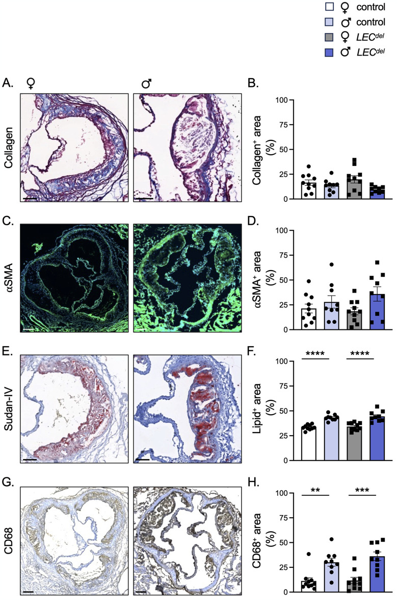
Fig 5. Reduced atherosclerotic plaque stability in male mice after 6 weeks of HCD.
Representative images and quantification of (immuno-) stainings for collagen (A,B; in blue), α-SMA (C,D; in green), Sudan-IV (E,F; in red) and CD68 (G,H; in brown) performed on aortic roots of female control Panx1fl/flApoe-/- mice (white), female Panx1LECdelApoe-/- mice (grey), male control Panx1fl/flApoe-/- mice (light blue) and male Panx1LECdelApoe-/- mice (dark blue) after 6 weeks of HCD. Scale bars represent 100 μm for A and E, and 200 μm for C and G. Mean ± SEM, N = 9–10, **P≤0.01, ***P≤0.001, ****P≤0.0001.

