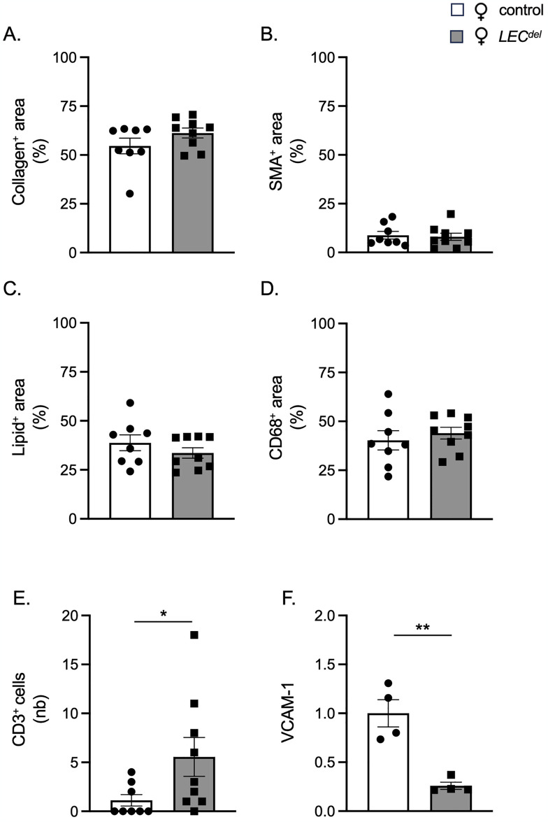
Fig 10. Panx1 deletion in LECs results in T cell enrichment in advanced atherosclerotic lesions of female mice.
Quantification of (immuno-)stainings for collagen (A), α-SMA (B), Sudan-IV (C), CD68 (D) and CD3 (E) in aortic roots of female Panx1fl/flApoe-/- mice (white) and female Panx1LECdelApoe-/- mice (grey) after 10 weeks of HCD. Mean ± SEM, N = 8–9. VCAM-1 expression (F) in Panx1-expressing (white) and Panx1-deficient (grey) LECs isolated from LNs of female mice. Mean ± SEM, n = 4. *P≤0.05, **P≤0.01.

