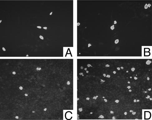
FIG. 8.
C. trachomatis serovars J and G infecting HeLa cells treated and untreated with cycloheximide. Low-magnification images of HeLa cells infected with C. trachomatis J/UW-36 (A and C) and serovar G/UW-57 (B and D). In each case, the cells were infected at a MOI of approximately 0.3 and fixed for microscopy 48 hpi. Cells shown in panels A and B were cultured in the presence of cycloheximide, while the cells shown in panel C and D were cultured in medium lacking cycloheximide. Each panel is labeled with antichlamydial LPS. Note that the absence of cycloheximide leads to a higher number of chlamydial inclusions in the cells infected with G/UW-57 but not in the cells infected with J/UW-36.

