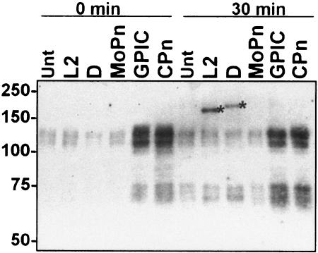
FIG. 6.
C. muridarum, C. caviae, and C. pneumoniae Tarp is not tyrosine phosphorylated. Total protein lysates were collected from HeLa cells that were either mock-infected (Unt) or infected (MOI, ∼100) with C. trachomatis L2 or D, C. muridarum (MoPn), C. caviae (GPIC), or C. pneumoniae (CPn) for 1 h at 4°C (time = 0) or at 30 min post-temperature shift to 37°C. Equal volumes of parallel mock-infected or infected cultures were loaded on each lane. Immunoblots were probed with 4G10 and visualized by chemiluminescence. Asterisks indicate the positions of tyrosine-phosphorylated Tarp; molecular masses in kilodaltons are indicated on the left.

