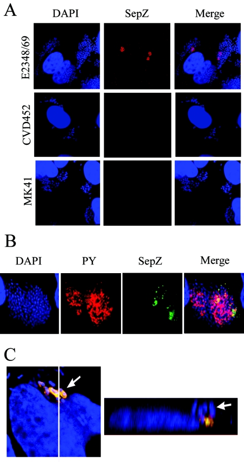
FIG. 6.
Localization of SepZ in infected HeLa cells by immunofluorescence confocal microscopy. (A) HeLa cells infected with E2348/69 (wild type), CVD451(escN), or MK41 (ΔsepZ) were colabeled with DAPI (blue) and polyclonal anti-SepZ antisera detected by AlexaFluor 568 goat anti-rabbit antibody (red). (B) Cells infected with E2348/69 were colabeled with DAPI (blue), monoclonal mouse anti-phosphotyrosine antibody was detected by AlexaFluor 568 goat anti-mouse antibody (PY; red), and polyclonal rabbit anti-SepZ antisera was detected with AlexaFluor 488 goat anti-rabbit antibody (green). The corresponding images of DNA, phosphotyrosine, and SepZ were combined (Merge). (C) Cells were infected with E2348/69 and stained identically to the cells shown in panel B. The image on the left represents 1 of 30 sequential z-series scans used to reconstruct the three-dimensional Z-section image on the right. The white line running from top to bottom in the left image denotes the location of the Z-section presented in the right image. White arrows point to the same bacterium in both images.

