FIG. 7.
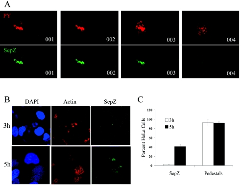
SepZ accumulation is detected in only a subset of pedestal regions after extended HeLa cell infections. (A) Four consecutive z-series scans of HeLa cells infected for 5 h with E2348/69 and colabeled with mouse anti-phosphotyrosine monoclonal antibody detected by AlexaFluor 568 goat anti-mouse antibody (PY; red) (top), and polyclonal rabbit anti-SepZ antisera detected with AlexaFluor 488 goat anti-rabbit antibody (Tir; green) (bottom). (B) HeLa cell monolayers infected for 3 or 5 h with E2348/69 and colabeled with DAPI (blue), AlexaFluor 568 phalloidin (Actin; red), and polyclonal rabbit anti-SepZ antisera detected with AlexaFluor 488 goat anti-rabbit antibody (green). (C) HeLa cells exhibiting actin-nucleation (pedestal formation) and/or SepZ accumulation after 3- and 5-h infections with E2348/69 were quantified. Cells from 10 fields at a magnification of ×100 (a minimum of 150 cells) were counted from each of three independent infections. The percentage of staining cells to total cells was calculated. The geometric means aregraphed, with standard deviation represented by error bars.

