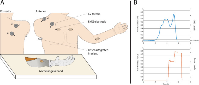
Fig. 6.
(A) The subject, seated in front of the Michelangelo hand, wearing the C2 tactors. The tactors were placed over soft tissue, avoiding the clavicle, coracoid process, and acromion of the scapula. The different placements of the EMG electrode used in this study are also shown. In the first two sessions, the electrode was placed over the right pectoralis muscle, while in sessions 3 and 4, it was positioned over the right and left pectoralis and the left flexor carpi radialis muscle. (B) The EMG and force traces the participant could see on the monitor during training. The horizontal lines are the thresholds for the different levels. The area below level 1 of EMG is the dead zone (see text).

