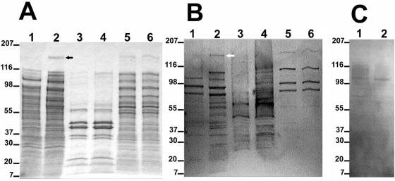
FIG. 5.
(A) SDS-PAGE of secreted, outer membrane, and cytoplasmic proteins stained with Coomassie blue. Lane 1, 10× concentrated supernatant of E. coli containing SuperCos1; lane 2, 10× concentrated supernatant of E. coli containing pCML76 (cfa clone); lane 3, outer membrane protein, E. coli containing SuperCos1; lane 4, outer membrane protein, E. coli containing pCML76; lane 5, cytoplasmic protein, E. coli containing SuperCos1; lane 6, cytoplasmic protein, E. coli containing pCML76. The numbers on the left indicate the positions of protein standards (in kilodaltons). The black arrow indicates the position of the 180-kDa protein. (B) Western blot transfer of panel A probed with a sera pool from 68 patients with cat scratch disease (IgG IFA titer ranging from 1:512 to 1:4,096 for B. henselae). The white arrow indicates the position of the 180-kDa immunogenic protein. (C) Western blot transfer of lanes 1 and 2 of panel A probed with patient serum negative for B. henselae antibodies (IgG IFA titer, <1:64).

