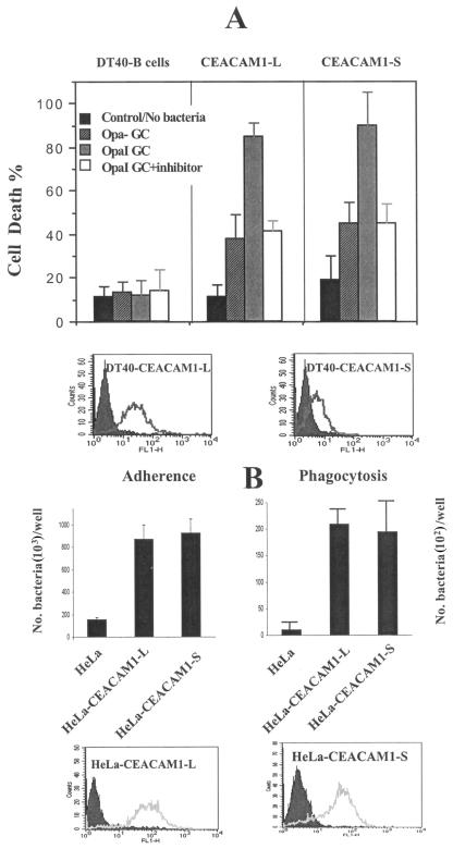
FIG. 3.
ITIM does not participate in CEACAM1-mediated the cell death and invasion. Both cDNAs of CEACAM1-long (L) and CEACAM1-short (S) forms were stably transfected into DT40 B cells (A) and HeLa cells (B). The expression levels of CEACAM1 in DT40 B-cell and HeLa cell transfectants were determined by flow cytometry using antibody YTH71.3, which recognizes CEACAM1, CEACAM6 (CD66c), and CEACAM3 (CD66d). Untransfected DT40 B or HeLa cells were used as a negative control (filled-in curve). DT40 or HeLa transfectants were challenged with Opa− and OpaI GC or E. coli and assayed for cell death (A) and phagocytic ability (B), respectively. The OpaI-mediated cell death of DT40-CEACAM1-L and -S cells was also tested in the presence of caspase-3 inhibitor (open bar, panel A). DT40 B cells and their transfectants that bound annexin V-FITC or took up PI were counted as dead cells. The percentage of dead cells is indicated on the y axis. CFU recovered after gentamicin treatment in HeLa cells were quantified as phagocytosed bacteria (B).

