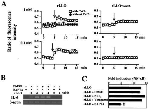
FIG. 4.
Involvement of Ca2+ in LLO-induced IL-6 expression. (A) Effect of rLLO on intracellular Ca2+. Caco-2 cells were treated with Fura 2-AM. Recombinant LLO or rLLOW492A was added to cells at the time indicated by the arrow. Intracellular Ca2+ was assessed by microfluorometry, and the results are expressed as the ratio of fluorescence intensity (510 nm). The y axis is (emission with 340-nm excitation)/(emission with 380-nm excitation). The results are representative of the results of three similar experiments. (B) Inhibition of rLLO-induced IL-6 expression by chelating intracellular Ca2+. Caco-2 cells were treated with 10 μM BAPTA-AM for 1 h and then stimulated with different doses of rLLO for 3 h. The expression of IL-6 was analyzed by RT-PCR. The results are representative of the results of three similar experiments. DMSO, dimethyl sulfoxide. (C) Inhibition of rLLO-induced NF-κB activation by chelating intracellular Ca2+. HEK293 cells were transfected with reporter vectors and treated with 10 μM BAPTA-AM, 50 μM NiCl2, and 10 μM verapamil for 1 h. The cells were then stimulated with 4 nM rLLO for 6 h, and NF-κB activation was determined by the luciferase assay. The results are expressed in units relative to the activity of the internal control pRL-SV40. The results are representative of the results of two similar experiments. The data are the means ± standard deviations for three determinations. Two asterisks indicate that there was a significant difference compared to cells stimulated with rLLO without any pretreatment.

