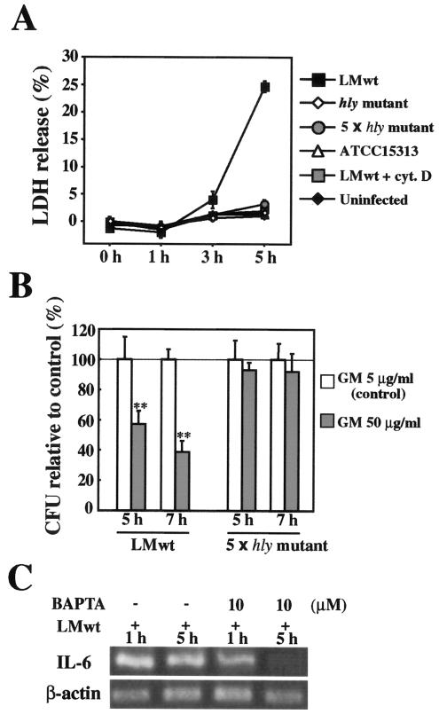
FIG. 5.
Involvement of Ca2+ in L monocytogenes-induced persistent IL-6 expression. (A) LDH release from L. monocytogenes-infected Caco-2 cells. Caco-2 cells were infected with the hly mutant, a fivefold-higher dose of the hly mutant (5 × hly mutant), ATCC 15313, or L. monocytogenes wt (LMwt) in the presence or absence of cytochalashin D (cyt. D). Culture supernatants were collected at different times, and the LDH activity in culture supernatants was assayed. The results are representative of the results of three similar experiments. The data are the means ± standard deviations for three determinations. (B) Gentamicin (GM) influx induced by L. monocytogenes infection. Caco-2 cells were infected with L. monocytogenes wt or a fivefold-higher dose of the hly mutant and incubated in medium containing 5 μg/ml (control) or 50 μg/ml of gentamicin for 5 and 7 h. The number of intracellular bacteria was determined by the CFU assay, and the results were expressed as the number of CFU relative to the control. The solid line indicates the value for the control. The results are representative of the results of two similar experiments. The data are the means ± standard deviations for three determinations. Two asterisks indicate that there was a significant difference compared to cells treated with 5 μg/ml gentamicin. (C) Inhibition of L. monocytogenes-induced persistent IL-6 expression by chelating intracellular Ca2+. Caco-2 cells were pretreated with BAPTA-AM for 1 h and then infected with L. monocytogenes wt for 1 or 5 h. Then the expression of IL-6 was analyzed by RT-PCR. The results are representative of the results of two similar experiments.

