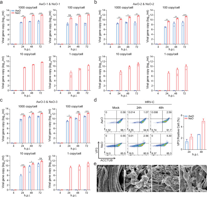
Fig. 4. Nasal organoids were more permissive to HRV-C than airway organoids.
a–c Airway (AwO) and nasal (NsO) organoids derived from different donors were inoculated in parallel with HRV-C3 at 1000, 100, 10, and 1 viral gene copy/cell (n = 3). Culture media were harvested from the infected organoids at the indicated h.p.i. to detect viral replication. d After co-staining with α-VP3 and α-ACCTUB, HRV-C3- and mock-infected airway and nasal organoids were applied to flow cytometry analysis (n = 2). Representative histograms are shown on the left. Data on the right represent the mean and SD from a representative experiment. e SEM images of HRV-C3- and mock-infected airway organoids. The experiment was independently performed three times with similar results. Scale bar, 200 nm. Data represent mean and SD of the indicated number (n) of biological replicates from a representative experiment. Statistical significance (in a, b, and c) was determined using a two-tailed Student’s t-test. *P < 0.05, **P < 0.01, ***P < 0.001. ns not significant. Source data are provided as a Source Data file for Fig. 4a–d.

