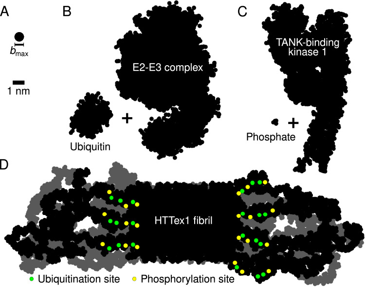
Fig. 8. PRD-domain brushes prevent post-translational-modification enzymes from reaching their target sites in HTTex1 fibrils.
A Size of the largest molecular species (= 0.8 nm) that can penetrate the polymer brush formed by the C-terminal PRD domains (as given by the polymer brush theory, see “Methods”). Silhouettes of the (B) E2-E3 Ubiquitin-Conjugating Complex (1C4Z) + ubiquitin (1UBQ), and (C) TANK-Binding Kinase 1 (6CQ0) + phosphate. D Silhouette of the HTTex1 fibril. The view is along the fibril growth direction. Shown are two consecutive monomer layers: the top layer in black, the layer behind it in gray. Post-translational modification sites in the top layer highlighted in color: the potential phosphorylation sites (residues T3, S13, and S16 in the N-terminal N17 domain) in yellow, and ubiquitination sites (residues K6, K9, and K15, also in the N17 domain) in green.

