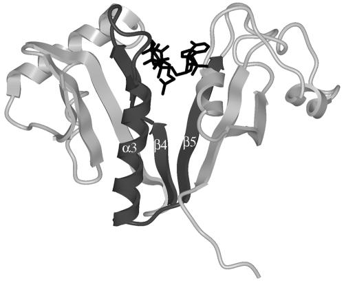
FIG. 2.
Structure of AAC(6′)-Ii (PDB 1B87) in complex with acetyl-CoA (23). The amino acids in motif A have been colored dark gray, and the acetyl-CoA molecule is shown in black. Note that the β4 strand is shown as two strands separated by a short coil (H74-P75) in this model.

