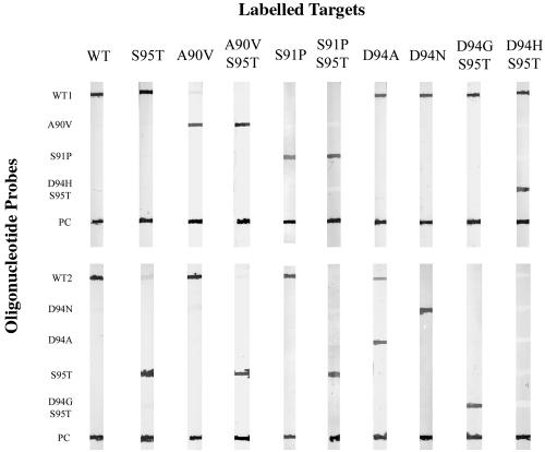
FIG. 2.
Representative line probe assay pattern obtained by testing the M. tuberculosis strains listed in Table 1. Oligonucleotide probes, indicated on the left, were blotted onto nitrocellulose strips and hybridized to wild-type and mutated DIG-labeled PCR product targets (top). Each strip was blotted with a wild-type DIG-labeled PCR product as a positive control for the development reaction (PC). PCR products from wild-type strains hybridized only to probes WT1 and WT2, while those containing the S95T polymorphism hybridized to the WT1 and S95T probes. Targets with single mutations in region 1 (A90V or S91P) hybridized to the specific probe and to the WT2 probe in the nonmutated region; when mutations were associated with the S95T polymorphism (A90V-S95T, S91P-S95T), hybridization occurred with the specific probe and with the S95T probe. Targets with a mutation in region 2 (D94A, D94N, D94G-S95T, or D94H-S95T) hybridized to WT1 and to the specific probes in region 2. Some cross-hybridization to the WT2 probe was observed with the D94A target.

