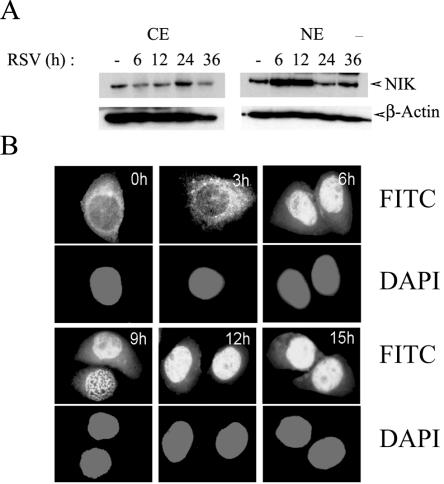
FIG. 6.
RSV-induced nuclear translocation of NIK. (A) Western immunoblot of 100 μg of NE or CE prepared from uninfected or RSV-infected A549 cells probed for endogenous NIK (top panel). The times of infection are shown at top (h). For the bottom panel, β-actin was probed as a loading control. Cytoplasmic NIK was depleted at 6 and 12 h with a concomitant increase in nuclear NIK. After 24 h, NIK redistributes back into the cytoplasm. (B) RSV-induced NIK nuclear translocation. Immunofluorescence microscopy was performed with anti-NIK antibody in a time course of RSV-infected A549 cells. The time (in hours) for each point is indicated at the top. In parallel, cells were stained with DAPI (4′,6′-diamidino-2-phenylindole) to localize nuclei. NIK is strongly nuclear 6 h after RSV infection. Western blotting and confocal microscopy were repeated twice with similar results.

