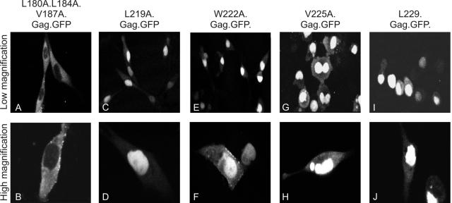
FIG. 2.
Identification of hydrophobic residues comprising the RSV Gag NES. The subcellular localization of mutant Gag-GFP fusion proteins was determined using confocal microscopy following transient transfection of QT6 cells. The amino acid substitutions for each mutant are shown above each panel. Top panels represent lower magnification images of Gag-GFP-expressing cells, while lower panels are higher-magnification images of adjacent fields.

