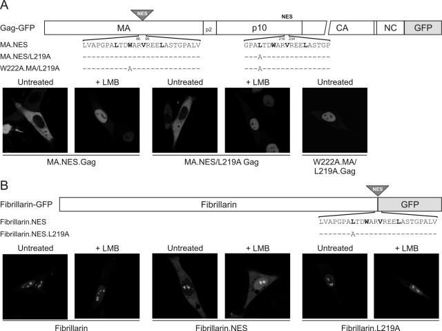
FIG. 6.
Mapping of a positionally independent and transferrable NES in Gag p10. (A) The position of the ectopic p10 NES sequence inserted into Gag-GFP is indicated by a shaded triangle, and the domains of Gag are indicated as MA, p2, p10, CA (capsid), NC, and PR (not drawn to scale). Residues 86 to 99 of MA were replaced with 216 to 234 of the p10 domain (plus three extra residues inserted with the restriction site introduced for cloning), and the amino acid sequences of the p10 NES and L219A and W222A mutants are shown below the diagram. Critical hydrophobic residues are indicated in bold type, and identical residues are represented as dashes. The subcellular localization of the MA.NES.Gag-GFP protein and NES variants was determined using confocal microscopy following transfection of QT6 cells that were either untreated (left panels) or incubated with 18 nM LMB (right panels). (B) Schematic diagram of fibrillarin-GFP showing the position of the inserted NES motif and the amino acid sequences of the p10 NES and the L219A mutant. QT6 cells expressing the indicated fibrillarin-derived GFP fusion proteins were untreated or treated with LMB and examined by confocal microscopy.

