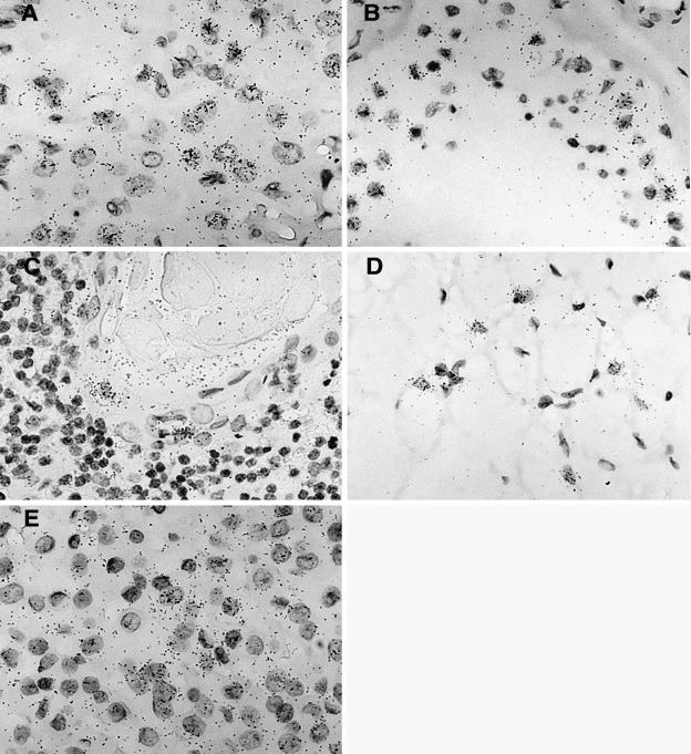
FIG. 5.
Hybridization signal obtained with the ERV3 antisense SU probe in five different tissues. (A) Tissue section from a corpus luteum demonstrating a positive and elevated ERV3 signal in progesterone-producing luteinized follicular cells. (B) Positive signal in yet unidentified cells obtained from a tissue section from a human, partly atrophic, testis with reduced spermatogenesis. (C) Tissue section from thymus with an elevated signal in cells in the outer part of a Hassal's body. (D) Brown fat section showing a high ERV3 message in typical multivacuolated brown fat cells. (E) Tissue section from a human pituitary gland (adenohypophysis portion), where an elevated ERV3 message is visible in all cell types.

