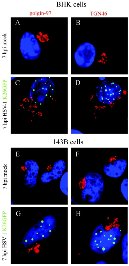
FIG.1.
Choice of cell line. To assess the disruption of the Golgi and TGN upon HSV-1 infection, BHK and 143B cells were mock treated or infected with HSV-1 K26GFP for 7 h at 37°C and labeled in parallel with golgin-97 (Golgi) or TGN46 (TGN) primary antibodies, followed with Alexa 568 secondary antibody. The samples were then examined by immunofluorescence (green, virus; red, Golgi or TGN). Comparison between BHK mock-infected cells (A and B) and BHK-infected cells (C and D) demonstrated a clear disruption of the Golgi and the TGN upon infection. In contrast, the morphology of the Golgi (E and G) and the TGN (F and H) were usually intact for 143B cells.

